


“I had more room and didn’t feel so trapped than in previous exams, and the scan went much faster. The visual guidance made me feel more confident despite my hearing issues.”
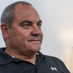
Francisco Javier Cubo, Oncological patient
Clínica Universidad de Navarra, Pamplona, Spain
