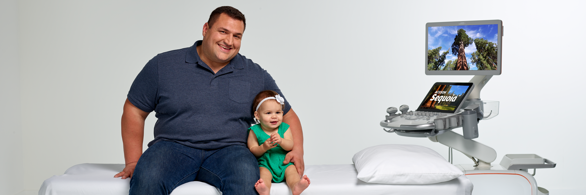
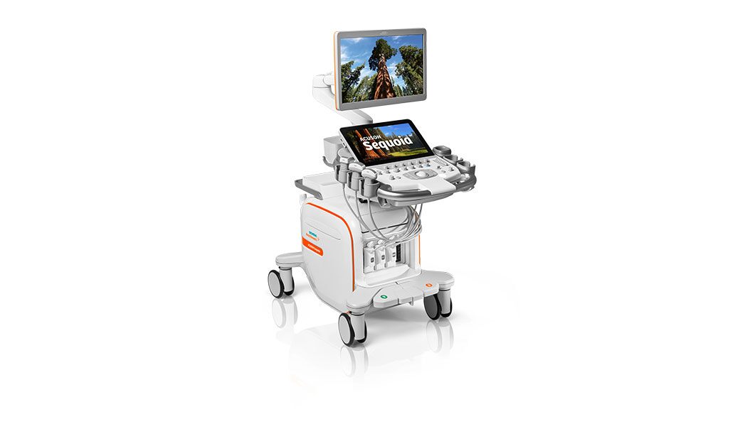
ACUSON Sequoia Ultrasound System
Taking Ultrasound to New Heights
The original ACUSON Sequoia™ is arguably the most popular ultrasound system we have ever created. In image quality, color sensitivity and advanced imaging modes, ACUSON Sequoia was – and still is an industry benchmark. The new ACUSON Sequoia is a remarkable evolution of a product that was so right in so many ways.
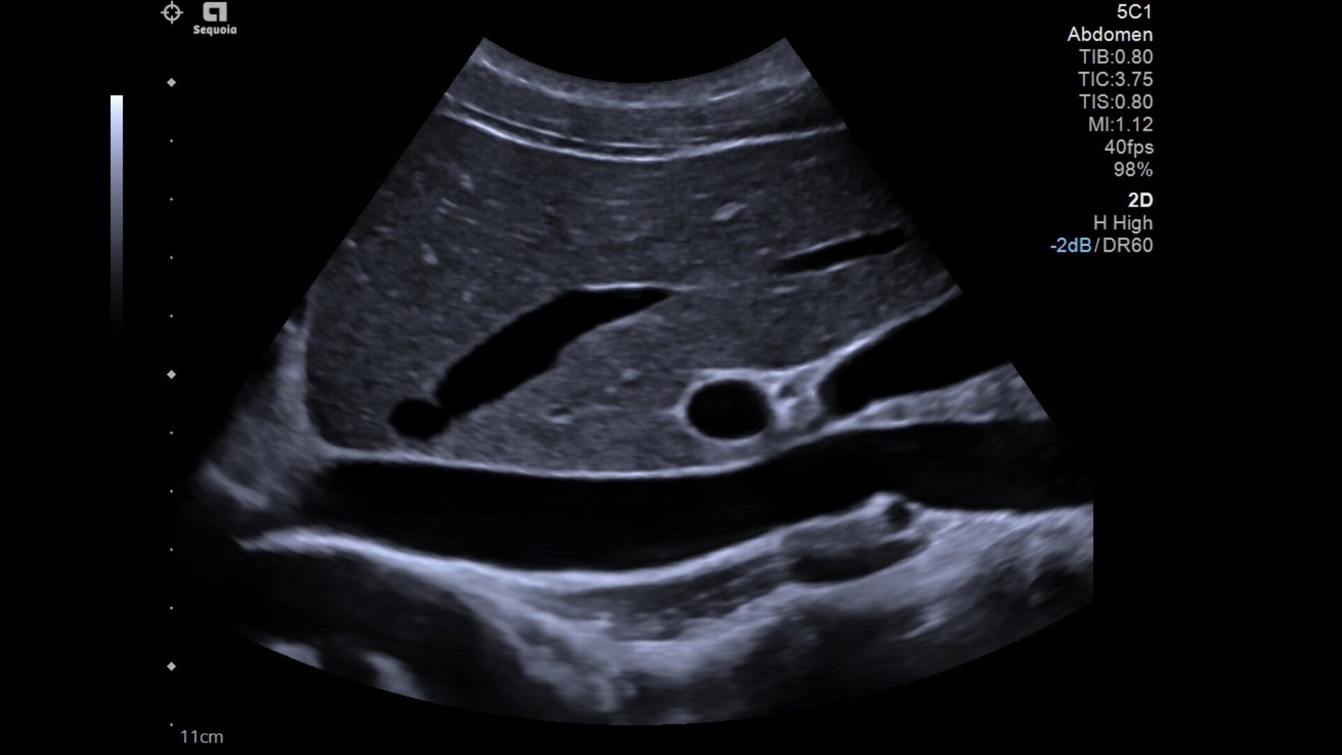
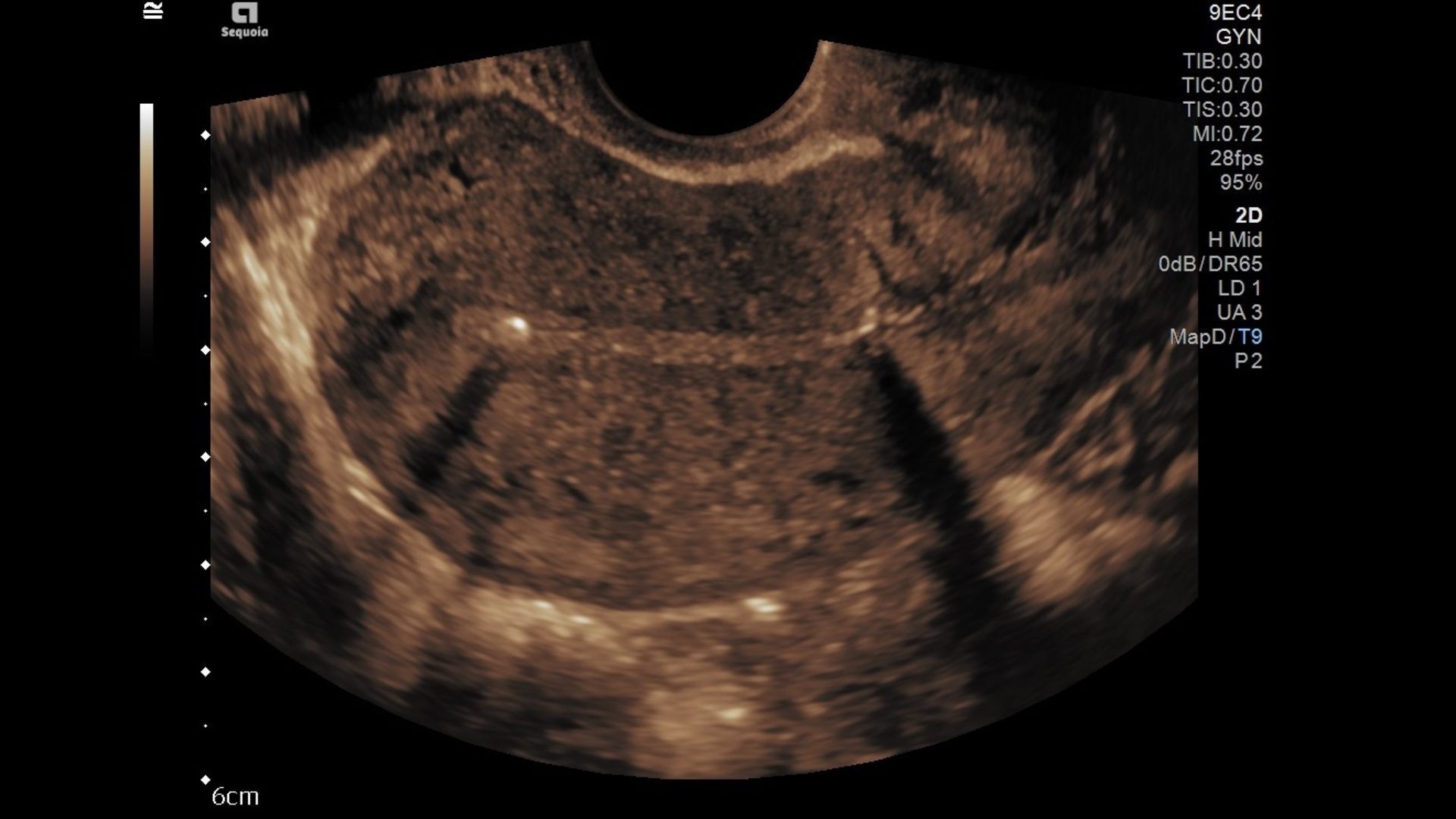
The first immediate benefit of the new ACUSON Sequoia is a remarkably fast, fully focused B-mode image without degradation of near-field or far-field resolution. We developed unique and patented technologies that allow ACUSON Sequoia to virtually eliminate color flash artifacts and penetrate deeper than conventional ultrasound systems.
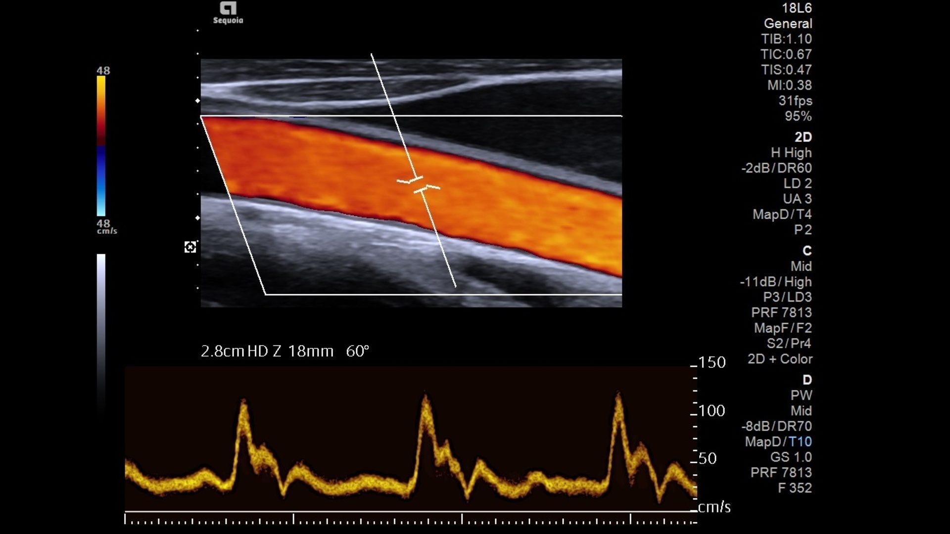
The second immediate benefit is color flow and color sensitivity without flashing or motion artifacts.
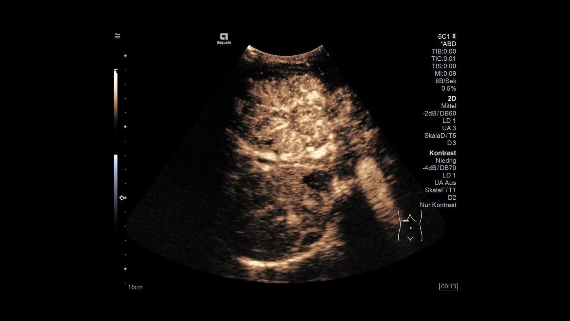
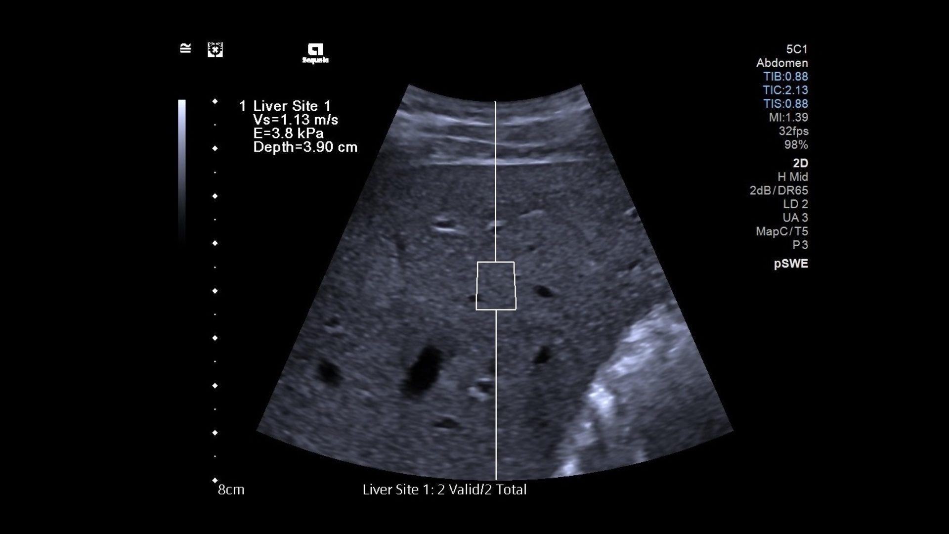
The third benefit comes in the form of deep tissue penetration without impacting diagnostic quality. Our team extensively researched a solution that would allow clinicians to image patients varying in size with confidence and clarity. From this research, a new transducer has emerged—DAX. Together with the ACUSON Sequoia we can for the first time deliver unprecedented images at depth without compromising diagnostic quality. DAX (Deep Abdominal Transducer) can penetrate as deep as 40cm.
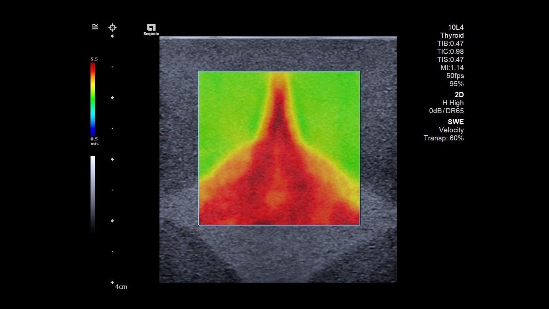
Ultrasound imaging is expected to deliver definitive and timely answers to important clinical questions. These answers must be provided in the most accurate and reproducible way. The new ACUSON Sequoia addresses these challenges with a comprehensive suite of advanced applications to deliver personalized ultrasound. With the power of the new ACUSON Sequoia, our elastography solution just got better.
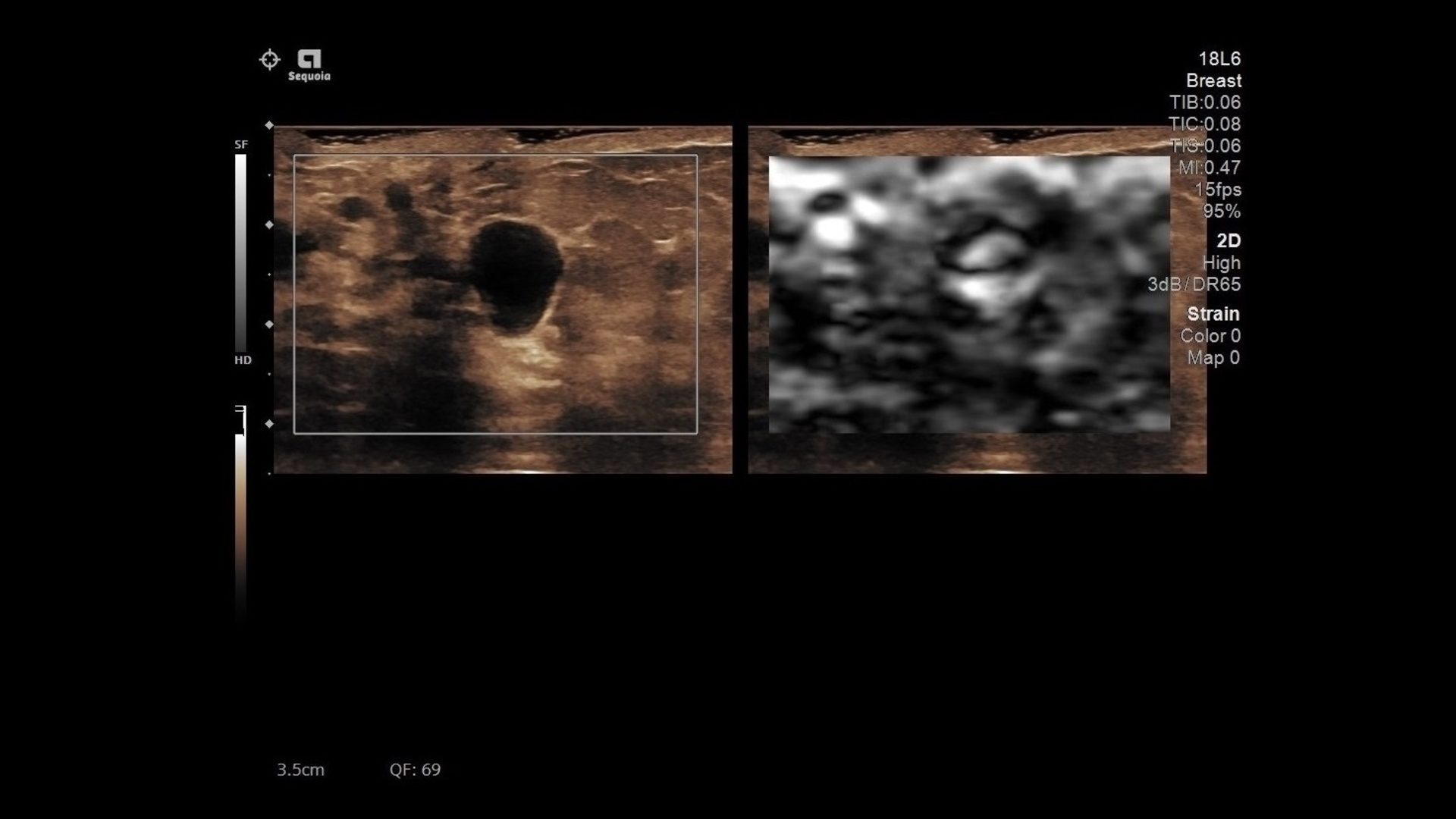
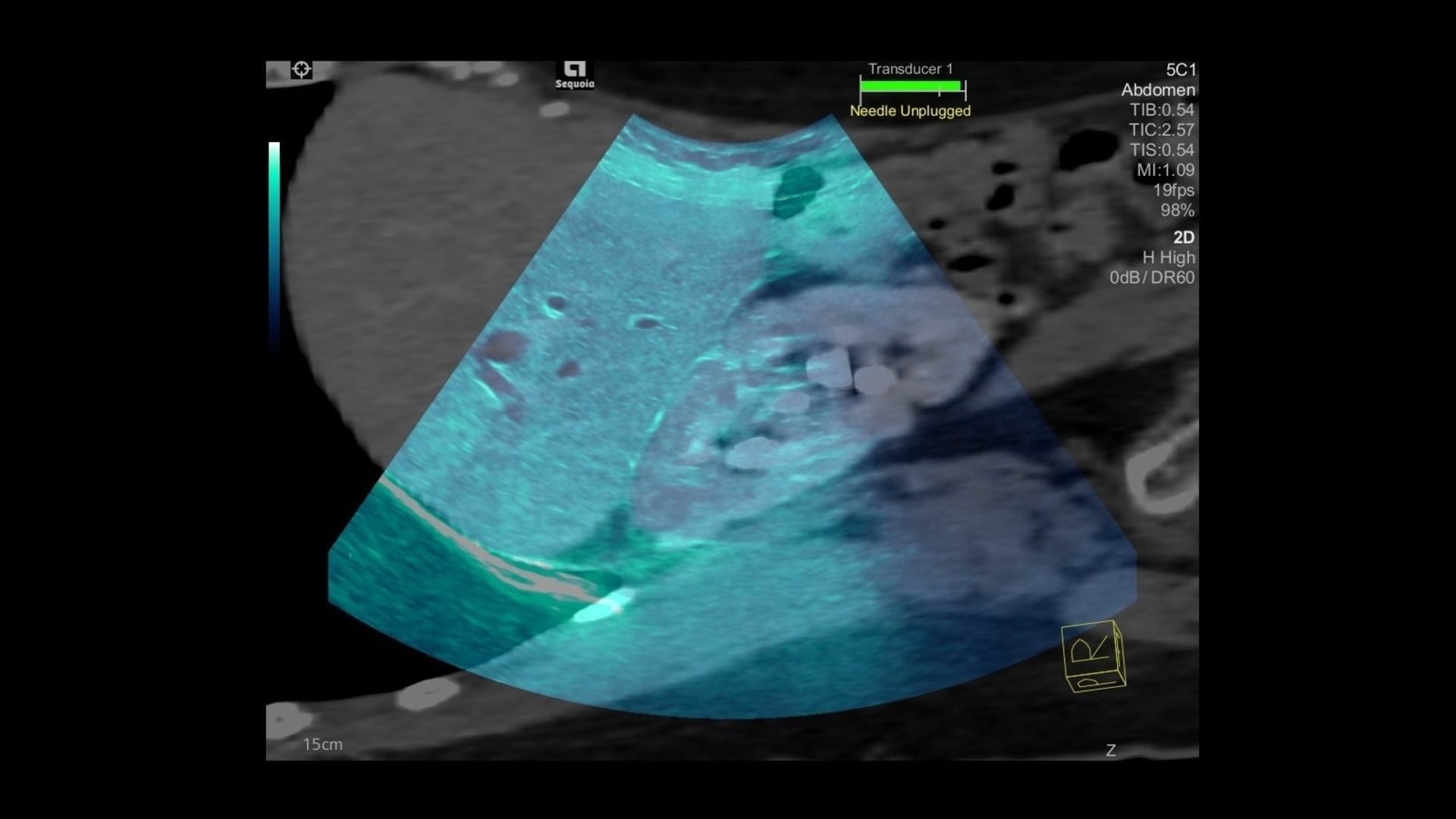
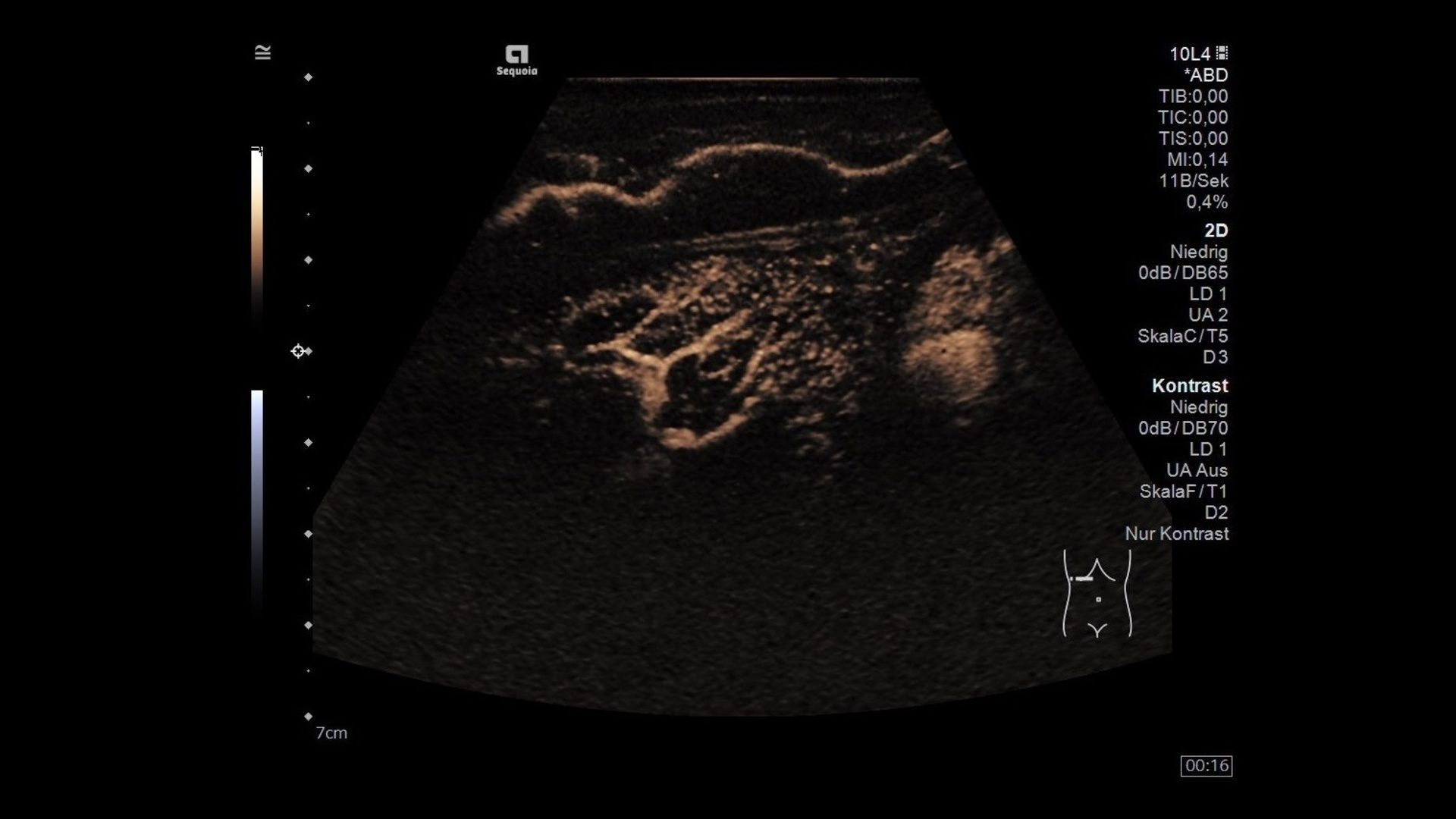
Contrast-enhanced ultrasound allows for detailed real time visualization of vessel morphology. With the new ACUSON Sequoia, the view time of contrast agents is significantly longer, allowing clinicians more time to scan for additional incidental lesions during their examinations and with up to twice the sensitivity.
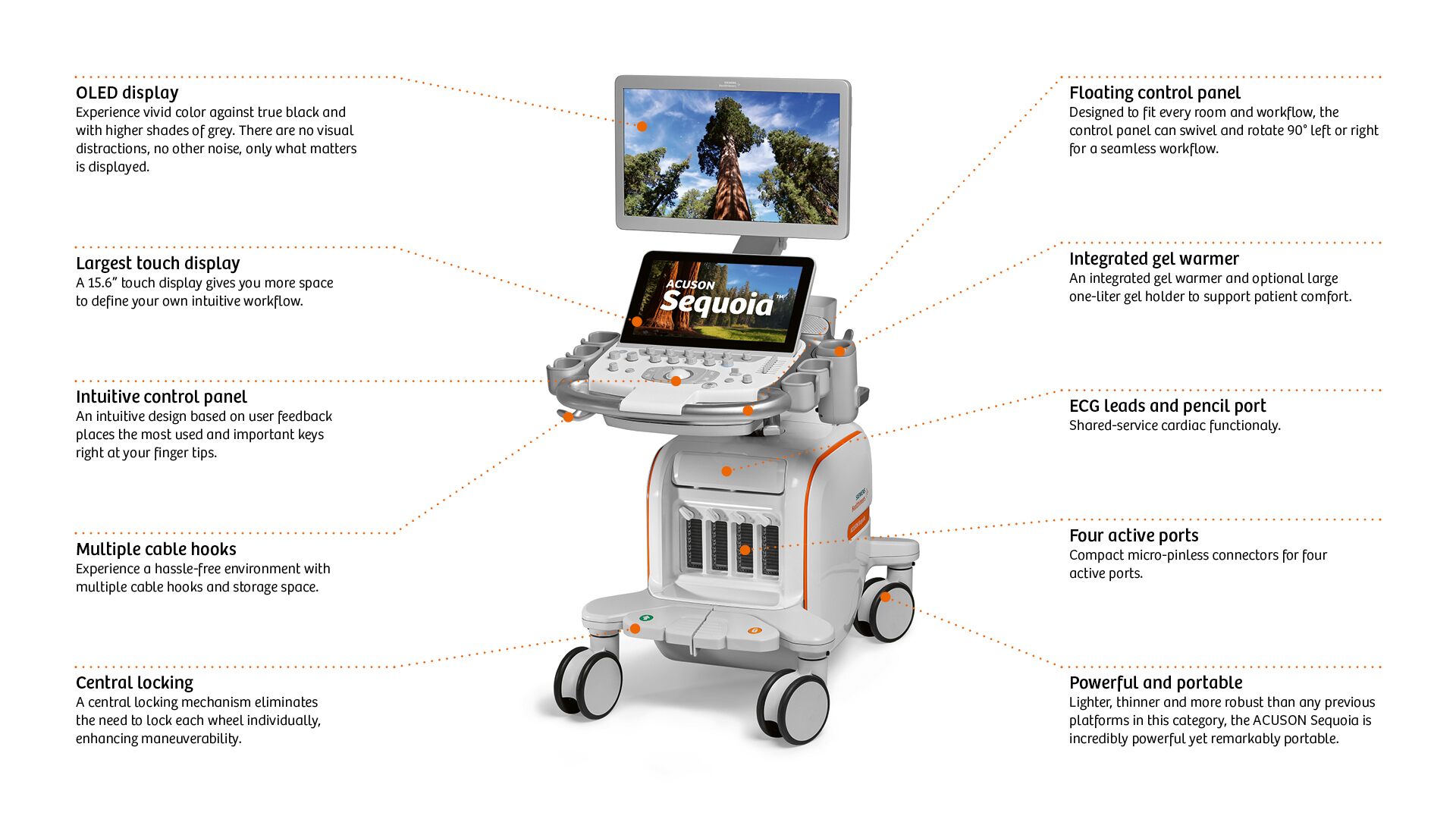
The variability inherent in the ultrasound scanning process can pose a challenge for the interpreting physicians. In an effort to eliminate variability, we hosted 170 workshops with 365 ultrasound users worldwide to create an ultrasound system designed by the user for the user.
With ACUSON Sequoia, each individual component is assembled to enable the system to accurately track the ultrasound signal throughout the signal path. From our power supplies to our receivers and graphics processing unit; to the compact-pinless transducer connectors to the transducer lens, we aimed to preserve the signal acoustic fidelity. This is all driven by the goal of accurately representing human biology. Siemens Healthineers calls this BioAcoustic imaging technology.